
 |
Case Report
A challenging case of encephalopathy in an endemic region of Texas with normal neuroimaging: A case report
1 Department of Internal Medicine, Ascension Seton Medical Center Austin, Austin, TX 78705, USA
2 Department of Internal Medicine, Summa Health System, Akron, OH 44304, USA
3 Department of Infectious Disease, Ascension Seton Medical Center Austin, Austin, TX 78705, USA
Address correspondence to:
Saketh Parsi
Department of Internal Medicine, Ascension Seton Medical Center Austin, Austin, TX 78705,
USA
Message to Corresponding Author
Article ID: 100024Z16SP2026
Access full text article on other devices

Access PDF of article on other devices


How to cite this article
Parsi S, Daggubati V, Subramany R, Ponnam HC, Bagwell K. A challenging case of encephalopathy in an endemic region of Texas with normal neuroimaging: A case report. J Case Rep Images Infect Dis 2026;9(2):10–15.ABSTRACT
Introduction: Murine typhus is a zoonotic disease caused by Rickettsia typhi, an obligate intracellular, gram-negative bacteria; in the past rats and their fleas have been the main reservoirs and vectors, but currently more cases are linked to cats, opossums, and cat fleas. The bacteria cause infection of the vascular endothelial cells, resulting in vasculitis, increased vascular permeability and fluid leakage, which may lead to pulmonary and cerebral edema. When immune cells in the visceral organs are infected, acute kidney injury, hepatitis, and encephalitis can occur. Neurological symptoms, particularly encephalitis, are uncommon. Blood tests generally reveal metabolic abnormalities including hypoalbuminemia, hypokalemia, raised inflammatory markers, elevated liver enzymes, and thrombocytopenia. A confirmatory diagnosis can be made by detecting high antibody titers together with a fourfold rise on repeat testing. The cerebrospinal fluid (CSF) usually has pleocytosis with the lymphocytes being the dominant cell type. Neuroimaging may vary from being normal to showing cerebral edema or infarction due to vasculitis.
Case Report: A 78-year-old woman was brought from another hospital because she had symptoms of generalized weakness and a change in mental state. Her initial laboratory tests indicated low potassium levels, low albumin levels, raised liver enzymes, and increased inflammatory markers. A chest computed tomography (CT) scan showed multifocal pneumonia. She was first given a broad-spectrum antibiotic treatment for the pneumonia; but since her encephalopathy did not improve, a consultation was arranged to consider the possibility of encephalitis. The results of the lumbar puncture were found to be consistent with a traumatic tap, and the cerebrospinal fluid analysis, together with the encephalitis panel, was otherwise normal. The neuroimaging did not show any acute abnormalities. Further information was obtained which showed that she had significant contact with cats, leading to a suspicion of murine typhus. The antibiotic treatment was changed to doxycycline. Serological tests confirmed the diagnosis, the antibody titer being 1:512. Her mental condition improved considerably as a result of doxycycline alone.
Conclusion: Murine typhus encephalitis should be considered as part of the differential diagnosis in cases of patients who have altered mental status and metabolic imbalances, especially in instances where both the cerebrospinal fluid and neuroimaging are normal, particularly in areas where the disease is endemic such as Texas. By taking a thorough history of the patient’s exposure and geographical background, including any contact with cats, it is possible to facilitate a timely diagnosis and suitable treatment. Clinical outcomes are better when the condition is detected and treated early.
Keywords: Cat fleas, Encephalitis, Endemic, Meningoencephalitis, Murine typhus, Rat fleas, Zoonotic infection
Introduction
Murine typhus (MT) is an acute febrile disease caused by Rickettsia typhi, a gram-negative coccobacillus which has to stay within host cells if it is to survive; the disease is mainly spread by infected rat fleas, rats serving as the main reservoirs [1]. Murine typhus is found naturally in the United States, particularly in coastal tropical and subtropical areas that have large populations of rodents. Recently, other zoonotic cycles have been reported which involve opossums and cats as hosts and cat fleas as vectors, especially in southern California, southern Texas, and Hawaii [2]. Although rarer globally, the disease can also occur in other tropical and subtropical coastal regions where climatic conditions support rodent hosts [3]. Rickettsial diseases are divided into typhus, spotted fever, transitional, and ancestral groups, each group being caused by a different species of Rickettsia. Unlike MT, which typically results in a mild fever, infections such as Rocky Mountain Spotted Fever and Epidemic Typhus can cause serious illness and death [4].
Murine typhus usually causes headache, muscle aches, joint aches, nausea, general discomfort, and fever, with the rash typically appearing after the fever starts. The rash usually first appears in the armpit area or on the inner arms before spreading to the trunk and the limbs [5]. R. typhi infects the vascular endothelium of small and medium-sized blood vessels, causing vasculitis and microvascular damage, which in turn increases vascular permeability and fluid to leak out, causing cerebral or pulmonary edema and can result in hypoalbuminemia, hypovolemia, and hyponatremia; these conditions in turn stimulate the release of antidiuretic hormone, causing hypokalemia and hypocalcemia. Additionally, the infection of monocytes and macrophages in organs including the lungs, liver, skin, heart, kidneys, and brain can give rise to myocarditis, pneumonitis, meningoencephalitis, and acute injury of the liver and kidneys [4].
The fact that MT encephalitis should be included in the differential diagnosis is shown by this case, even though it is rare, in particular when a patient has altered mental status, metabolic disturbances, a history of cat exposure, negative results from the standard encephalitis panel, no abnormalities on imaging, and is from an endemic area such as Texas.
Case Report
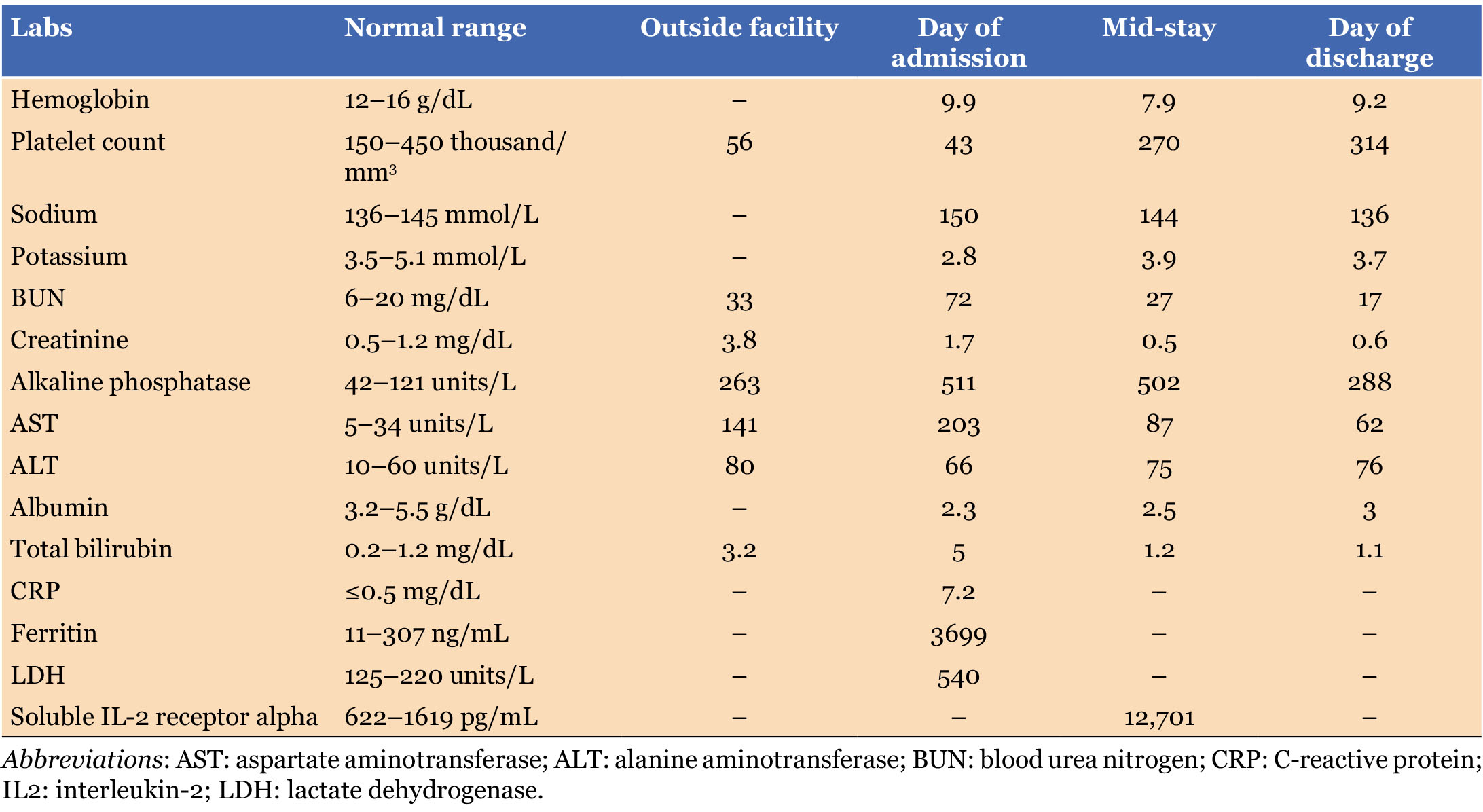
A 78-year-old female with no important medical history initially went to another hospital where she was admitted for general weakness and had fallen; she was then transferred to our hospital since she had an altered mental state which might have been caused by a seizure. Blood tests at the outside hospital showed thrombocytopenia, with a platelet count of 56,000/mm3, and elevated blood urea nitrogen (BUN) 33 mg/dL, creatinine 3.8 mg/dL, and other abnormalities such as anion gap 23, total bilirubin 3.2 mg/dL, alkaline phosphate 263 U/L, aspartate aminotransferase (AST) 141 U/L, and alanine aminotransferase (ALT) 80 U/L. Computed tomography (CT) of the abdomen was unremarkable, and CT angiography of the head and neck showed no intracranial or extracranial stenosis, hemorrhage, or infarction. She was given meropenem as an empirical treatment for possible sepsis caused by a urinary tract infection, and had one unit of platelets and five units of cryoprecipitate administered to her before she was transferred.
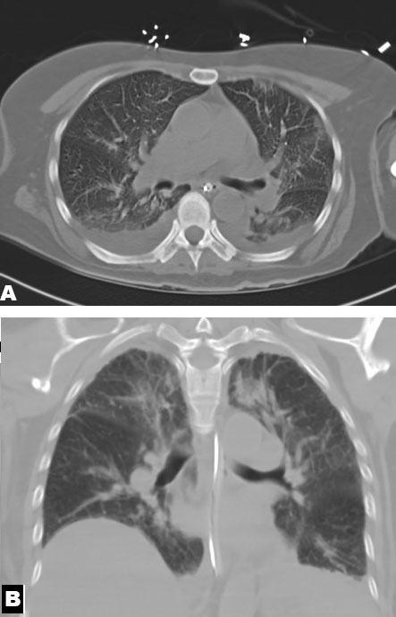
When the patient got to our hospital, she was still confused and opened her eyes only to verbal or painful stimulation, failing to obey commands or to stay attentive. The repeat blood tests showed that thrombocytopenia was still present, the platelet count being 43,000/mm3, hemoglobin 9.9 g/dL, sodium 150 mmol/L, BUN 72 mg/dL, creatinine 1.7 mg/dL, alkaline phosphate at 511 U/L, AST 203 U/L, ALT 66 U/L, total bilirubin 5 mg/dL, direct bilirubin 2.7 mg/dL, potassium 2.8 mmol/L, calcium 7.8 mg/dL, and albumin 2.3 g/dL (Table 1). The urine analysis was normal and did not show any infection. The chest CT demonstrated multifocal pneumonia with small bilateral pleural effusions (Figure 1A and Figure 1B). An ultrasound of the abdomen and the kidneys was carried out in order to investigate the transaminitis and the acute kidney injury, and it revealed diffuse fatty infiltration of the liver and a gallbladder that was absent. The patient was started on intravenous (IV) cefepime, one gram every 12 hours, and azithromycin 250 mg, dosed according to kidney injury and considering pneumonia as the cause of encephalopathy. Also, the patient was started on levetiracetam 500 mg twice daily for possible seizures.
The patient’s mental status worsened despite antibiotic treatment, leading to consultations with neurology and infectious disease (ID) specialists, considering meningoencephalitis as the cause. Additional blood tests showed no schistocytes in the peripheral blood smear; ammonia 42 µmol/L; ferritin 3699 ng/mL; C-reactive protein (CRP) 7.2 mg/dL; soluble interleukin-2 receptor alpha 12701 pg/mL; and lactate dehydrogenase 540 U/L. An extensive infectious and autoimmune workup was unrevealing, with negative results for hepatitis panel, rapid plasma reagin (RPR), HIV antigen/antibody, anti-double-stranded DNA, anti-Jo-1, centromere B antibodies, RNP antibodies, SCL-70 antibodies, and Sjögren’s SSA/SSB antibodies. However, a positive antinuclear antibody at 1:160 in a speckled pattern, which is not specific in acute infection. Hematology evaluation ruled out thrombotic thrombocytopenic purpura due to the absence of schistocytes and ruled out Hemophagocytic lymphohistiocytosis, as it did not meet five of the eight HLH-2004 criteria. Infectious disease and neurology services recommended electroencephalography (EEG), brain magnetic resonance imaging (MRI) with and without contrast, lumbar puncture, and cerebrospinal fluid (CSF) studies.
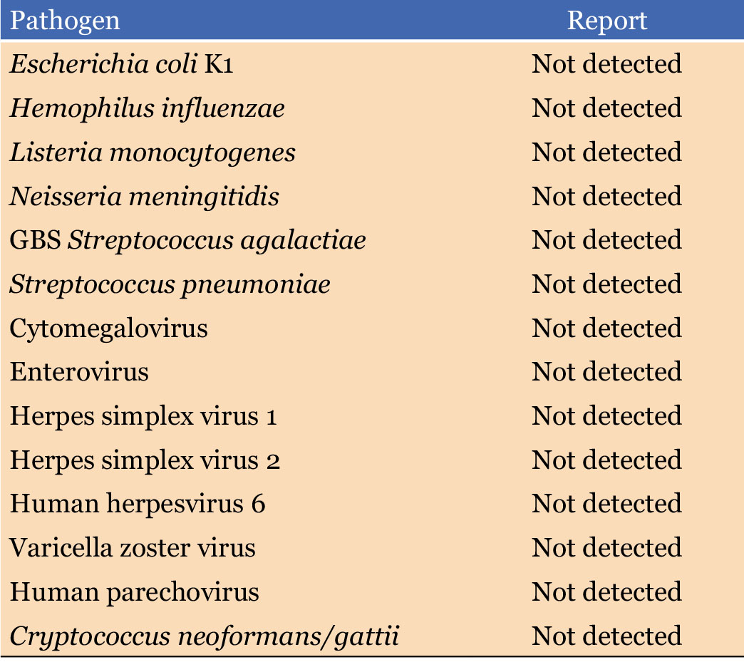
Cerebrospinal fluid analysis (CSF) showed a total nucleated cell count of 15/mm3, red blood cells 350/mm3, glucose 56 mg/dL, and protein 105 mg/dL. Cerebrospinal fluid testing showed positive varicella zoster virus (VZV) IgG 2.04, while VZV IgM was negative at 0.07. CSF West Nile virus IgG and IgM, CSF cultures, and the CSF encephalitis panel were all negative (Table 2). Brain MRI demonstrated no acute abnormalities, and long-term video EEG revealed severe diffuse cerebral dysfunction without epileptiform discharges. Empiric intravenous acyclovir was initiated for possible viral meningitis but was later discontinued after serum and CSF VZV polymerase chain reaction (PCR) testing returned negative despite positive CSF VZV IgG. Additional history obtained from the patient’s daughter revealed that the patient was highly functional at baseline and routinely cared for both domestic and wild cats. Given the combination of thrombocytopenia, elevated liver enzymes, encephalopathy, and cat exposure, azithromycin was transitioned to IV doxycycline 100 mg twice daily for suspected murine typhus. Infectious disease consultants noted that, although uncommon, murine typhus can present with meningoencephalitis. Murine typhus serology subsequently demonstrated an IgG titer of 1:512 five days later. The patient completed the Cefepime course for multifocal pneumonia.
Neurology expressed concern regarding discontinuation of acyclovir because of the patient’s persistent altered mental status; however, the decision was deferred to the infectious disease specialist. Infectious disease continued holding acyclovir, given negative serum and CSF VZV PCR results, and concluded that the isolated CSF VZV IgG positivity likely reflected passive transfer from a traumatic lumbar puncture in the setting of reactive serum VZV IgG and negative IgM (<0.91). A repeat murine typhus IgG titer was ordered to evaluate for a fourfold rise to further support the diagnosis, but the reported titer was unchanged at 1:512. Nevertheless, the patient demonstrated significant clinical improvement in mental status while receiving doxycycline monotherapy, strongly supporting murine typhus-associated meningoencephalitis as the underlying etiology. She completed a 14-day course of doxycycline during hospitalization and was discharged home on levetiracetam.
Discussion
This instance shows an uncommon way in which murine typhus can present itself as meningoencephalitis. Murine typhus is found in many parts of the world and is generally limited to tropical and subtropical coastal regions where rats and cats act as hosts and the rat and cat fleas function as vectors for the transmission of R. typhi [1]. In the United States, MT should be included in the differential diagnosis of patients who present with symptoms of meningoencephalitis, such as altered mental status and seizures, especially in endemic areas like southern California, Texas, and Hawaii [2]. The fact that our patient lived in Texas and had contact with cats highlights the need to take a detailed medical history, covering the patient’s geographic location and any exposure to animals.
About 2% of people who have MT develop meningitis or meningoencephalitis, which raises the case fatality rate to 27%. Headache is the most frequent neurological symptom observed in cases of murine typhus, affecting around 81% of patients, photophobia (10%), confusion or delirium (8%), stupor (4%), seizures (4%), and ataxia (1%) [4]. In the present case, the patient was transferred to us from another hospital due to general malaise. When she arrived at our facility, she was unable to speak and had reduced alertness.
The laboratory results in the case of MT show low platelet count, low hemoglobin level, leukopenia or relative lymphocytosis, low albumin level, hypokalemia, hyponatremia, increased GGT and alkaline phosphatase, increased creatinine, and elevated lactate dehydrogenase (LDH). Moreover, typhus is confirmed by immunoglobulin M and G antibody titers >1:150 and >1:400, respectively, and by a fourfold increase in IgG titers between the acute and convalescent phases, which occur 2–10 weeks after the first sample [6]. Other diagnostic modalities, such as R. typhi DNA PCR, have high specificity but low sensitivity, because their diagnostic yield depends on bacterial load and the timing of illness. PCR is most useful during the acute phase, particularly within the first week of infection. For this reason, serologic testing using antibody titers is generally more appropriate [7]. In our case, the patient had most of the metabolic derangements described in the case report and also had elevated murine typhus titers. However, the confirmation lab result was similar to the previous titers, 1:512, which could mean either that the patient was already in the convalescent stage when presented or that the patient is not yet in the convalescent phase, as this can occur anytime between 2 and 10 weeks.
Spears et al. [8] reported that CSF analysis showed at least one of the following abnormalities: pleocytosis, elevated protein concentration, or decreased glucose levels. Furthermore, CSF leukocytosis was predominantly lymphocytic. Elevated CSF protein was observed in 78.8% of patients, and hypoglycorrhachia was present in 33.3%. In our case, CSF analysis revealed a TNC count of 15 cells/mm3, elevated protein, and a normal glucose level; however, the findings were inconclusive due to the potential confounding effect of a traumatic lumbar puncture, as evidenced by an RBC count of 350/mm3.
Neuroimaging findings in murine typhus encephalitis, head CT findings range from normal studies to evidence of cerebral edema or cerebral infarction. Similarly, brain MRI findings range from normal to abnormalities such as cerebral edema, cerebral infarction, or leptomeningeal enhancement [8],[9]. In our case, the head CT and brain MRI findings were reported as unremarkable on radiologic interpretation. From the therapeutic standpoint, doxycycline is the treatment of choice for murine typhus and, when started promptly, is associated with defervescence within 48–72 hours in most patients. Alternative agents include macrolides (e.g., azithromycin), fluoroquinolones (e.g., ciprofloxacin), and chloramphenicol, although the latter is no longer available for clinical use in the United States [4].
Conclusion
Neurological signs of murine typhus are rare, with encephalitis being especially uncommon. However, MT should still be considered in patients with an altered mental status, particularly those with a history of exposure to cats or living in endemic areas like Texas, California, or Hawaii. Our case was challenging to diagnose because neuroimaging appeared normal and cerebrospinal fluid analysis yielded few abnormalities, but likely traumatic. Still, careful assessment of metabolic imbalances, thorough history-taking regarding exposures and residence, and close collaboration with specialists ultimately led to the diagnosis of murine typhus encephalitis. The patients’ cat exposure was a key epidemiologic clue that raised suspicion. Early detection and proper treatment played a critical role in the patient’s recovery and positive outcome.
REFERENCES
1.
Riswari SF, Prodjosoewojo S, Mony SR, Megantara I, Iskandar S, Mayasari W, et al. Murine typhus is a common cause of acute febrile illness in Bandung, Indonesia. PLoS One 2023;18(7):e0283135. [CrossRef]
[Pubmed]

2.
Faccini-Martínez ÁA, Walker DH, Blanton LS. Murine typhus in Latin America: Perspectives of a once recognized but now neglected vector-borne disease. Am J Trop Med Hyg 2022;107(4):740–6. [CrossRef]
[Pubmed]
3.
Snellgrove AN, Goddard J. Murine typhus: A reemerging rickettsial zoonotic disease. J Vector Ecol 2024;50(1):1–13. [CrossRef]
[Pubmed]
4.
Blanton LS. Murine typhus: A review of a reemerging flea-borne rickettsiosis with potential for neurologic manifestations and sequalae. Infect Dis Rep 2023;15(6):700–16. [CrossRef]
[Pubmed]
5.
Dye BV, Coba JA, Dayton CL, Cadena J, Anstead GM. Flea-borne typhus as a COVID-19 mimic: A report of four cases. Case Rep Infect Dis 2024;2024:9914306. [CrossRef]
[Pubmed]
6.
Dhawan S, Robinson MT, Stenos J, Graves SR, Wangrangsimakul T, Newton PN, et al. Selection of diagnostic cutoffs for murine typhus IgM and IgG immunofluorescence assay: A systematic review. Am J Trop Med Hyg 2020;103(1):55–63. [CrossRef]
[Pubmed]
7.
Lokida D, Sudarmono P, Kosasih H, Butar-Butar DP, Salim G, Antonjaya U, et al. Comparison of commercial enzyme-linked immunosorbent assay and immunofluorescence assay for diagnosis of acute Rickettsia typhi infections. Vector Borne Zoonotic Dis 2020;20(2):93–9. [CrossRef]
[Pubmed]
8.
Spears CE, Chandramohan D, Tanabe MB, Anstead NC, Turkily ELM, Blanton LS, et al. Meningoencephalitis in flea-borne typhus: A report of two cases and a review of CNS complications. Pathogens 2026;15(6):590. [CrossRef]
[Pubmed]
9.
Loayza Pintado J, Aboytes J, Uribe C. A flea-borne mystery: Unraveling murine typhus in a patient with unexplained encephalopathy. J Investig Med High Impact Case Rep 2025;13:23247096251345086. [CrossRef]
[Pubmed]
SUPPORTING INFORMATION
Acknowledgments
Grammarly is the platform used only for spell checks and grammar corrections.
Author ContributionsSaketh Parsi - Conception of the work, Design of the work, Acquisition of data, Drafting the work, Revising the work critically for important intellectual content, Final approval of the version to be published, Agree to be accountable for all aspects of the work in ensuring that questions related to the accuracy or integrity of any part of the work are appropriately investigated and resolved.
Vinoop Daggubati - Conception of the work, Design of the work, Drafting the work, Final approval of the version to be published, Agree to be accountable for all aspects of the work in ensuring that questions related to the accuracy or integrity of any part of the work are appropriately investigated and resolved.
Revathy Subramany - Conception of the work, Design of the work, Drafting the work, Final approval of the version to be published, Agree to be accountable for all aspects of the work in ensuring that questions related to the accuracy or integrity of any part of the work are appropriately investigated and resolved.
Harikrishna C Ponnam - Conception of the work, Design of the work, Drafting the work, Final approval of the version to be published, Agree to be accountable for all aspects of the work in ensuring that questions related to the accuracy or integrity of any part of the work are appropriately investigated and resolved.
Kelli Bagwell - Conception of the work, Design of the work, Drafting the work, Final approval of the version to be published, Agree to be accountable for all aspects of the work in ensuring that questions related to the accuracy or integrity of any part of the work are appropriately investigated and resolved.
Guaranter of SubmissionThe corresponding author is the guarantor of submission.
Source of SupportNone
Consent StatementWritten informed consent was obtained from the patient for publication of this article.
Data AvailabilityAll relevant data are within the paper and its Supporting Information files.
Conflict of InterestAuthors declare no conflict of interest.
Copyright© 2026 Saketh Parsi et al. This article is distributed under the terms of Creative Commons Attribution License which permits unrestricted use, distribution and reproduction in any medium provided the original author(s) and original publisher are properly credited. Please see the copyright policy on the journal website for more information.

