|
Case Report
Persistent fever and focal tibial swelling after laboratory-confirmed dengue revealing acute hematogenous tibial osteomyelitis in a toddler: A diagnostic challenge in a resource-limited setting
1 MBChB, MMed Paediatrics, Department of Pediatrics, Gargaar Multispecialty Hospital, Hargeisa, Somaliland
2 MBChB, MCS Tropical Pediatrics, Department of Pediatrics, Gargaar Multispecialty Hospital, Hargeisa, Somaliland
Address correspondence to:
Noradin Garad Egeh Elmi
MBChB, MMed Paediatrics, Department of Pediatrics, Gargaar Multispecialty Hospital, Hargeisa,
Somaliland
Message to Corresponding Author
Article ID: 100023Z16NE2026
Access full text article on other devices

Access PDF of article on other devices


How to cite this article
Elmi NGE, Ali MM. Persistent fever and focal tibial swelling after laboratory-confirmed dengue revealing acute hematogenous tibial osteomyelitis in a toddler: A diagnostic challenge in a resource-limited setting. J Case Rep Images Infect Dis 2026;9(2):1–9.ABSTRACT
Introduction: Dengue fever is common in tropical and subtropical regions and may present with fever, rash, lethargy, and generalized musculoskeletal symptoms. However, persistent fever accompanied by progressive focal musculoskeletal findings should prompt reassessment for an additional invasive bacterial process.
Case Report: A 19-month-old boy with laboratory-confirmed dengue initially received supportive outpatient management. Approximately 10 days later, he developed persistent high-grade fever, progressive swelling, and tenderness over the right tibia, limping, and refusal to bear weight. Laboratory investigations demonstrated marked leukocytosis with granulocytic predominance, C-reactive protein (CRP) of 111 mg/L, and erythrocyte sedimentation rate of 70 mm/hour. Plain radiography showed subtle anterior tibial soft-tissue changes, while targeted ultrasonography reportedly demonstrated periosteal thickening. Acute hematogenous tibial osteomyelitis was considered the most likely clinical diagnosis. Empirical intravenous ceftriaxone-sulbactam produced no clear clinical improvement after 48 hours, and therapy was changed to intravenous vancomycin. The child subsequently became afebrile, local inflammatory findings resolved, limb function returned, and CRP decreased to <5 mg/L. He was discharged on oral cephalexin to complete therapy. Microbiological confirmation and magnetic resonance imaging (MRI) were unavailable.
Conclusion: Laboratory-confirmed dengue should not preclude diagnostic reassessment when persistent fever is accompanied by new focal musculoskeletal findings. In resource-limited settings, repeated clinical examination, serial inflammatory markers, and available imaging may support timely recognition and management of probable bacterial osteomyelitis when microbiological confirmation and advanced imaging are unavailable.
Keywords: Acute hematogenous osteomyelitis, Bacterial infection, Dengue, Resource-limited setting
Introduction
Dengue is one of the most common mosquito-borne viral infections worldwide and a major cause of childhood morbidity in tropical and subtropical regions. An estimated 390 million dengue virus infections occur annually, with approximately 96 million resulting in clinically apparent disease [1]. In children, dengue commonly presents with fever, rash, anorexia, lethargy, vomiting, and diffuse musculoskeletal symptoms, many of which are nonspecific and overlap with numerous other infectious conditions [2],[3]. While most children recover with supportive management, the broad clinical spectrum of dengue may complicate recognition of an unrelated but concurrent bacterial infection.
Acute hematogenous osteomyelitis (AHO) is an uncommon but potentially limb-threatening and life-threatening bacterial infection in children that requires prompt diagnosis and initiation of antimicrobial therapy to prevent irreversible bone destruction, growth disturbance, and other complications [4],[5]. Young children often present with nonspecific symptoms, and localized signs may be subtle or delayed, particularly during the early stages of disease [6]. Consequently, delayed diagnosis remains common, especially in resource-limited settings where advanced imaging and microbiological investigations are not readily available [7].
Although bacterial coinfections have been reported in patients with dengue, they are uncommon and encompass a broad spectrum of invasive infections rather than a disease-specific association [8],[9],[10]. Therefore, laboratory confirmation of dengue should not conclude the diagnostic evaluation when the subsequent clinical course is atypical. Persistent or recurrent fever, focal musculoskeletal findings, neutrophilic leukocytosis, and markedly elevated inflammatory markers are discordant with uncomplicated dengue and should prompt systematic reassessment for invasive bacterial disease, including acute hematogenous osteomyelitis, regardless of the initial viral diagnosis [11],[12].
Here, we report a 19-month-old boy with laboratory-confirmed dengue who subsequently developed acute hematogenous tibial osteomyelitis in a resource-limited setting where blood cultures, bone sampling, and magnetic resonance imaging (MRI) were unavailable. Rather than suggesting a unique dengue-associated syndrome, this case highlights the importance of diagnostic reassessment when the clinical course becomes atypical and demonstrates how careful serial clinical assessment, inflammatory markers, plain radiography, and targeted ultrasonography can support timely recognition and management of invasive bacterial disease when definitive diagnostic resources are limited.
Case Report
Patient information and initial illness
A 19-month-old boy was initially managed as an outpatient for laboratory-confirmed dengue based on positive NS1 antigen and dengue immunoglobulin M (IgM) tests. His illness began with high-grade fever, poor appetite, lethargy, and a generalized rash, consistent with uncomplicated dengue. He was previously healthy but underweight, with a history of eczema and incomplete immunization. There was no history of chronic medical illness, recurrent invasive infections, previous hospitalization, trauma, falls, intramuscular injections, skin wounds, cellulitis, or antibiotic exposure before the current illness.
Evolution of focal musculoskeletal findings
Approximately 10 days after the onset of dengue symptoms, the child’s clinical course changed. Rather than improving, he developed persistent fever associated with progressively worsening swelling over the middle-to-distal aspect of the right tibia, accompanied by warmth, erythema, marked localized tenderness, guarding, and crying on manipulation of the affected limb. He progressively reduced spontaneous movement of the leg, developed a limp, and ultimately refused to bear weight. Before the current illness, he had achieved normal independent walking and had no preceding gait abnormality. There was no clinical evidence of knee involvement.
Clinical examination
On admission, the child weighed 10 kg and appeared toxic and lethargic but remained hemodynamically stable without clinical evidence of dehydration. His temperature was 40.0°C, heart rate 150 beats/min, respiratory rate 33 breaths/min, blood pressure 90/50 mmHg, and oxygen saturation 99% while breathing room air.
Examination of the right lower limb demonstrated diffuse swelling over the middle-to-distal tibia with overlying warmth and erythema. Marked focal bony tenderness was elicited along the tibial shaft, and spontaneous use of the limb was significantly reduced because of pain. The overlying skin was intact, with no puncture wounds, abrasions, or cellulitis. Examination of the ipsilateral knee and ankle revealed no joint swelling, erythema, effusion, or restriction of passive range of motion, making septic arthritis less likely. No other focal abnormalities were identified on systemic examination.
Investigations
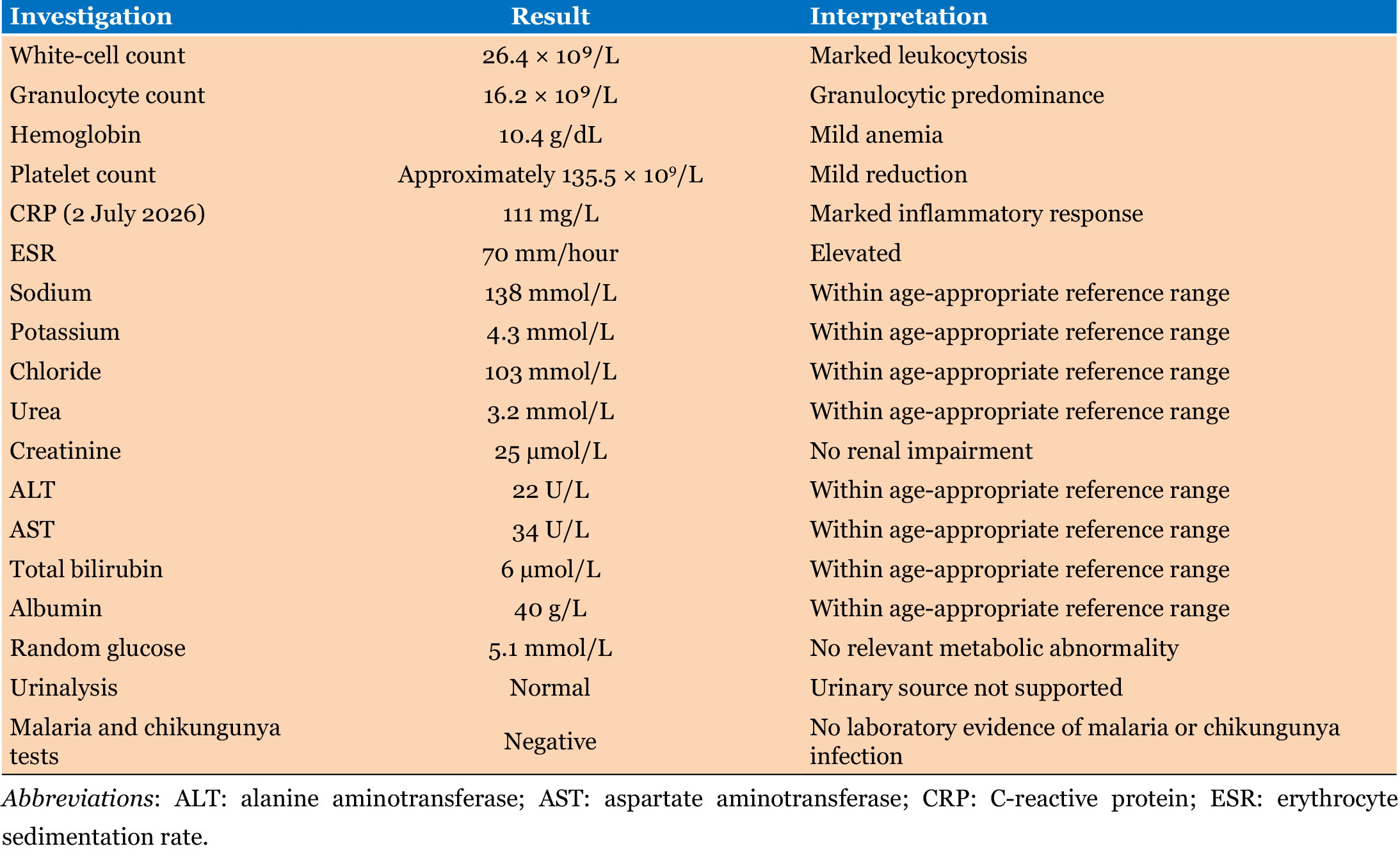
The laboratory findings at diagnostic reassessment are summarized in Table 1.
The persistence of high fever beyond the expected course of uncomplicated dengue, together with progressive focal tibial swelling, marked bony tenderness, refusal to bear weight, neutrophilic leukocytosis (white blood cell count 26.4 × 10⁹/L), CRP 111 mg/L, and erythrocyte sedimentation rate (ESR) 70 mm/hour, represented findings that were discordant with uncomplicated dengue. These clinical and laboratory abnormalities prompted reassessment for an additional diagnosis, with particular concern for an invasive bacterial musculoskeletal infection.
Blood culture facilities were unavailable at the treating institution. No blood cultures, bone aspirate, subperiosteal aspirate, or soft-tissue specimens were obtained, and antimicrobial susceptibility testing could not be performed. Consequently, microbiological confirmation of the causative organism was not possible.
Imaging
Plain radiographs of the right leg (29 June 2026) demonstrated subtle anterior tibial soft-tissue swelling without fracture, focal lytic lesions, periosteal reaction, sclerosis, or cortical destruction. Targeted ultrasonography demonstrated periosteal thickening over the affected tibia. Magnetic resonance imaging (MRI), the preferred modality for confirming osteomyelitis and defining disease extent, was unavailable. Although the imaging findings were nonspecific, when interpreted alongside the evolving focal examination findings and markedly elevated inflammatory markers, they supported a working clinical diagnosis of acute hematogenous osteomyelitis of the right tibia.
Differential diagnosis
The differential diagnosis included acute hematogenous osteomyelitis, septic arthritis, pyomyositis, cellulitis, occult trauma, dengue-associated musculoskeletal pain, tuberculous osteomyelitis, and malignancy.
Acute hematogenous osteomyelitis was considered the most likely diagnosis because of the combination of persistent fever, progressive focal bony swelling and tenderness, refusal to bear weight, marked neutrophilic leukocytosis, and substantially elevated CRP and ESR. Septic arthritis was considered less likely because the knee remained clinically unaffected, with no joint swelling, effusion, or restriction of passive movement. Occult trauma was not supported by the clinical history or radiographic findings, while pyomyositis and cellulitis were considered less likely because tenderness was localized to the tibia rather than the surrounding soft tissues. Tuberculous osteomyelitis and malignancy were considered less probable given the acute presentation, high inflammatory response, and rapid clinical improvement following antimicrobial therapy. Although microbiological confirmation was unavailable, the overall clinical, laboratory, and imaging findings were most consistent with acute hematogenous osteomyelitis.
Treatment and outcome
Empirical intravenous ceftriaxone-sulbactam (500 mg every 12 hours) was initiated because of suspected bacterial osteoarticular infection. After 48 hours of therapy, the child remained persistently febrile, with ongoing tibial swelling, marked tenderness, and refusal to bear weight, indicating an inadequate clinical response.
Given the persistent fever, progressive focal musculoskeletal findings, and markedly elevated inflammatory markers, antimicrobial therapy was revised to intravenous vancomycin at 15 mg/kg/dose (150 mg) every 8 hours. Following this change, the child demonstrated progressive clinical improvement, with resolution of fever, reduction in local swelling, warmth, and tenderness, and gradual restoration of spontaneous limb movement and weight bearing. Renal function remained normal throughout treatment.
After five days of intravenous vancomycin, the CRP had declined to <5 mg/L and the complete blood count had normalized. The child was discharged on 9 July 2026 weighing 9.6 kg and was prescribed oral cephalexin [25 mg/kg/dose (250 mg) every 8 hours] for an additional three weeks to complete therapy. The caregiver reported good adherence to treatment.
Following discharge, the child was reviewed in person approximately one week later and again at three weeks. At the first follow-up, he remained afebrile, with continued improvement in tibial swelling and tenderness and progressive restoration of weight bearing and right lower-limb use. At the three-week review, the local inflammatory signs had resolved, and he was walking normally with full use of the affected limb and no apparent gait abnormality or functional limitation. No recurrence of fever, tibial swelling, or focal tenderness was identified during these in-person assessments.
A further in-person review was scheduled for approximately six weeks after discharge; however, the family did not attend the scheduled appointment. The parents were subsequently contacted by telephone and reported that the child remained clinically well, was walking normally, and had experienced no recurrence of fever, tibial swelling, tenderness, or limitation of limb use. Repeat ultrasonography had been considered to assess interval resolution of the previously reported periosteal abnormality; however, it was not performed because the family was unable to meet the out-of-pocket cost of repeat imaging. Although the sustained clinical and biochemical improvement supported the working diagnosis of bacterial osteomyelitis and a favorable treatment response, the absence of microbiological confirmation precluded identification of the causative organism or determination of its antimicrobial susceptibility.
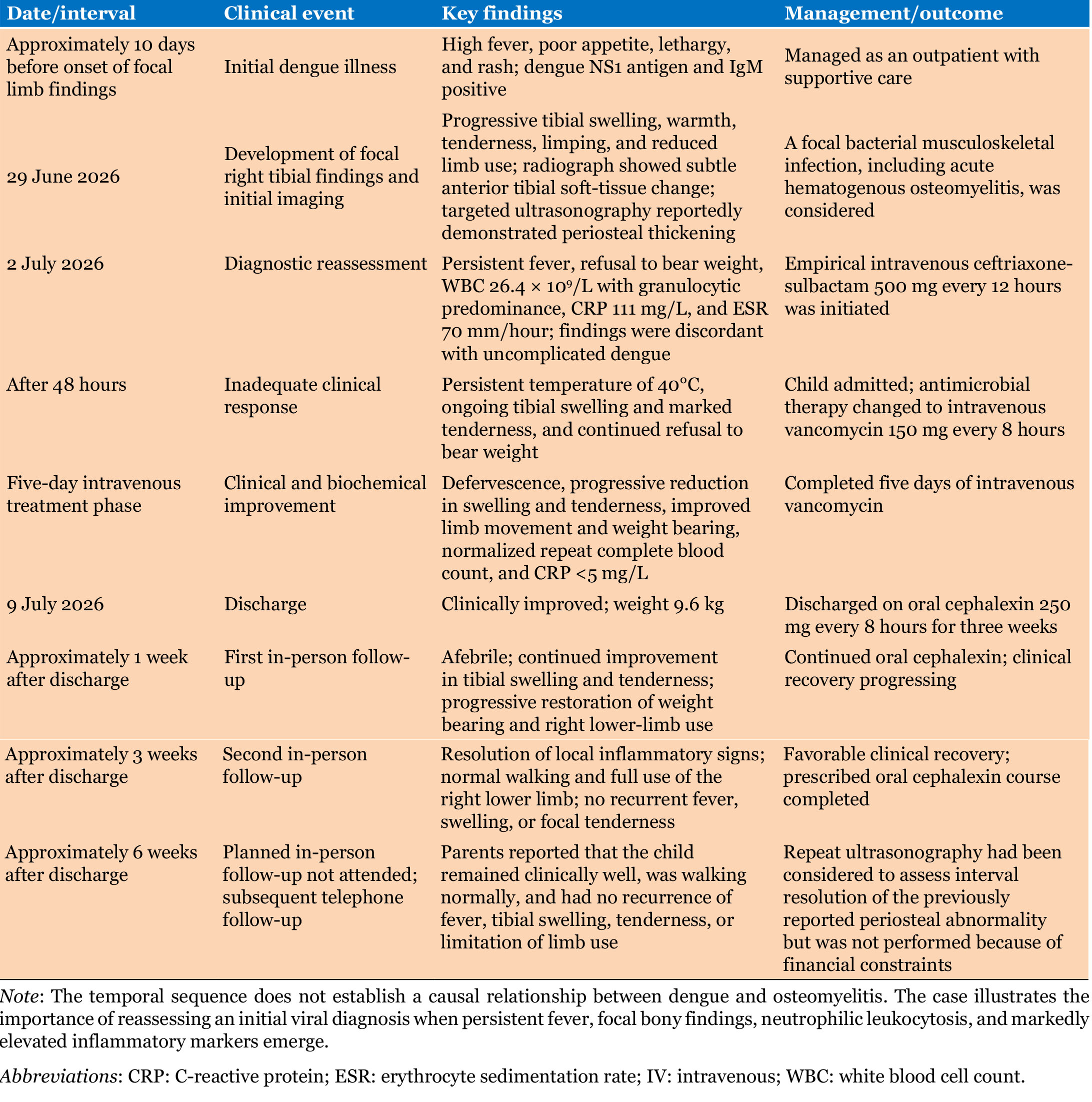
The chronological evolution of the illness, investigations, treatment, clinical response, and follow-up is summarized in Table 2.
Discussion
Diagnostic reassessment after laboratory-confirmed dengue
The central clinical lesson from this case is that laboratory confirmation of dengue should not terminate diagnostic evaluation when the subsequent clinical course becomes atypical. Dengue commonly causes fever, rash, lethargy, reduced appetite, and generalized musculoskeletal discomfort. In a preverbal child, reduced limb movement or reluctance to walk may therefore initially be attributed to viral myalgia. However, persistent high fever, progressive localized tibial swelling and tenderness, refusal to bear weight, granulocytic leukocytosis, and markedly elevated inflammatory markers were discordant with uncomplicated dengue and indicated the need to investigate an additional invasive bacterial process.
Published reports have described bacterial infections occurring during or shortly after dengue, including bacteremia, pneumonia, pyomyositis, septic arthritis, and osteomyelitis, with Staphylococcus aureus identified in several microbiologically confirmed cases [8],[9],[10]. These reports do not establish a distinct dengue-associated osteomyelitis syndrome. Rather, they illustrate that a confirmed viral diagnosis does not exclude concurrent or subsequent bacterial infection, particularly when fever persists, focal findings emerge, or inflammatory markers are substantially elevated.
Role of inflammatory markers
Inflammatory markers contributed to both diagnostic reassessment and monitoring of treatment response. At reassessment, the child had a white blood cell (WBC) count of 26.4 × 10⁹/L with granulocytic predominance, CRP of 111 mg/L, and ESR of 70 mm/hour. No single CRP or ESR threshold confirms acute hematogenous osteomyelitis, and values vary according to disease severity, timing, causative organism, bacteremia, and the presence of abscess formation. Nevertheless, the combination of markedly elevated inflammatory markers and focal bony abnormalities was much more suggestive of invasive bacterial infection than uncomplicated dengue.
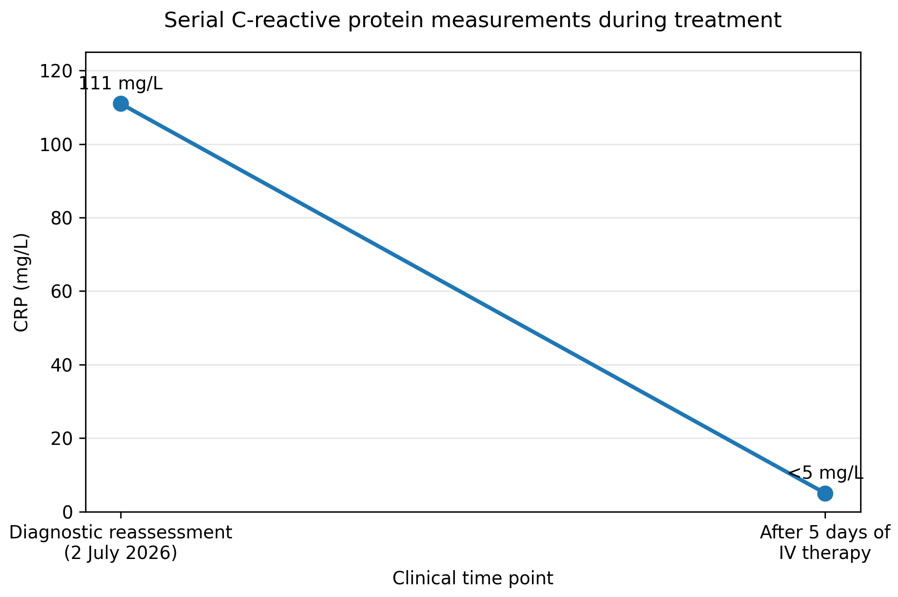
Serial CRP measurement was particularly useful because the concentration declined from 111 to <5 mg/L in parallel with defervescence, reduction in swelling and tenderness, improved limb use, and normalization of the complete blood count (Figure 1).
Imaging in a resource-limited setting
Plain radiographs have limited sensitivity during the early stages of acute osteomyelitis because cortical destruction, periosteal reaction, and other osseous abnormalities may not yet be visible. Their principal early value is to identify soft-tissue swelling and exclude fracture, tumor, foreign body, or another alternative diagnosis [5],[6],[7]. The subtle anterior tibial soft-tissue change observed in this child was therefore compatible with early infection but was not diagnostic.
Magnetic resonance imaging is the preferred advanced imaging modality because it can define marrow involvement, subperiosteal or intramuscular collections, joint extension, and the anatomical extent of disease [5],[6],[7].. It was unavailable in this setting. Ultrasonography cannot exclude intramedullary infection and should not be considered a substitute for MRI; however, it may demonstrate periosteal elevation or thickening, adjacent soft-tissue edema, subperiosteal fluid, or a drainable collection. In this case, targeted ultrasonography of the symptomatic right tibial region was performed during the initial assessment of the focal swelling and tenderness by an experienced sonographer as part of routine clinical imaging. The documented sonographic finding was periosteal thickening over the symptomatic tibial region. This finding was considered supportive of a deeper osteoarticular process when interpreted together with the marked focal tibial tenderness, swelling, refusal to bear weight, and substantially elevated inflammatory markers; however, ultrasonography was not considered diagnostic of osteomyelitis in isolation. Detailed technical parameters of the examination were not documented, and the original ultrasound images were not retained for retrospective review or inclusion in this report.
Antimicrobial management and diagnostic uncertainty
Empirical ceftriaxone-sulbactam was initially administered for suspected bacterial osteoarticular infection. After 48 hours, the child remained febrile with persistent tibial swelling, marked tenderness, and refusal to bear weight. The absence of clear improvement did not establish antimicrobial resistance. Because blood culture, bone sampling, susceptibility testing, and MRI were unavailable, it was not possible to determine whether the limited early response reflected antimicrobial coverage, insufficient treatment duration, an undetected collection, or another disease-related factor.
Alternative diagnoses were considered during reassessment. Cellulitis was considered because of the localized swelling, warmth, and erythema; however, the marked focal tenderness over the tibia, refusal to bear weight, substantially elevated inflammatory markers, and reported periosteal thickening on ultrasonography favored a deeper osteoarticular process rather than isolated superficial cellulitis. Pyomyositis was also considered given the fever and localized inflammatory findings; however, no focal intramuscular collection was reported on targeted ultrasonography, and the maximal tenderness was localized over the tibia rather than predominantly within the surrounding muscle. Occult trauma was considered because of the focal limb pain and refusal to bear weight; however, there was no reported history of trauma, and plain radiography did not demonstrate a fracture or other traumatic osseous abnormality. Although these findings favored acute hematogenous tibial osteomyelitis, the absence of MRI and microbiological confirmation meant that alternative diagnoses could not be excluded with absolute certainty.
Antimicrobial therapy was subsequently changed to vancomycin, after which the child showed progressive clinical and biochemical improvement. This temporal response supported the working diagnosis of bacterial osteomyelitis but did not identify the causative organism or prove methicillin-resistant Staphylococcus aureus (MRSA). The case should therefore be described as culture-unconfirmed acute hematogenous tibial osteomyelitis.
Intravenous-to-oral transition
Historically, pediatric acute hematogenous osteomyelitis was treated with prolonged intravenous therapy. Contemporary evidence supports transition to an active oral agent when the child is clinically improving, fever has resolved or is clearly resolving, oral medication can be tolerated, and inflammatory markers demonstrate a favorable trend [5]. This approach reduces hospitalization, intravenous-line complications, and cost without compromising outcomes in appropriately selected uncomplicated cases.
After five days of intravenous vancomycin, the child was afebrile, local swelling and tenderness had improved, limb use had returned, the repeat complete blood count had normalized, and CRP had decreased to <5 mg/L. He was therefore transitioned to oral cephalexin at 75 mg/kg/day in three divided doses for three weeks. The total planned treatment duration was approximately 26 days. Because the causative organism and antimicrobial susceptibility were unknown, close clinical follow-up was important after oral narrowing.
Comparison with published reports
The published literature remains too limited and heterogeneous to define a specific syndrome of osteomyelitis associated with dengue. Previously reported pediatric and adult cases have included staphylococcal bacteremia, septic arthritis, osteomyelitis, pyomyositis, and pneumonia during or shortly after dengue [8],[9],[10]. Compared with these reports, the present patient was a younger child with apparently isolated tibial disease who lacked microbiological and MRI confirmation because of resource limitations and recovered without documented surgical intervention.
The literature comparison should therefore be presented as contextual evidence that invasive bacterial infection can coexist with or follow dengue, rather than evidence that dengue directly caused the osteomyelitis.
Implications for resource-limited settings
This case reflects diagnostic constraints encountered in many dengue-endemic and resource-limited settings, where blood cultures, bone aspiration, MRI, pediatric orthopedic services, and therapeutic drug monitoring may be unavailable or financially inaccessible. These limitations reduce diagnostic certainty, restrict organism-directed therapy, and make it more difficult to identify abscesses requiring drainage.
When advanced investigations are unavailable, repeated bedside examination, serial inflammatory markers, careful interpretation of plain radiography, and targeted ultrasonography can support timely clinical decision-making. However, these approaches do not replace microbiological sampling or MRI, and uncertainty regarding the organism, antimicrobial susceptibility, and anatomical extent of infection must be reported transparently.
Strengths and limitations
The strengths of this report include laboratory-confirmed dengue, clearly documented evolution of focal musculoskeletal abnormalities, objective inflammatory-marker monitoring, recognition of an inadequate early clinical response, and documented clinical and biochemical recovery following treatment modification. The report also provides evidence from a pediatric setting that is underrepresented in the published literature.
The principal limitations are the absence of blood culture, bone or subperiosteal sampling, antimicrobial susceptibility testing, MRI, and original ultrasound images. Consequently, the causative organism and its antimicrobial susceptibility could not be determined, MRSA infection could not be confirmed, and the anatomical extent of osteomyelitis could not be fully characterized. The temporal relationship between dengue and osteomyelitis does not establish causation.
Follow-up was also limited: although the child was reviewed in person approximately one week and three weeks after discharge, with sustained clinical recovery and restoration of normal right lower-limb function, the family did not attend the planned six-week in-person review. Subsequent telephone contact with the parents indicated that the child remained clinically well, was walking normally, and had experienced no recurrence of fever, tibial swelling, or tenderness. Repeat ultrasonography had been considered to assess interval resolution of the previously reported periosteal abnormality but was not performed because of financial constraints. Therefore, although the available clinical and biochemical findings supported a favorable treatment response, longer-term resolution could not be confirmed by direct clinical examination or repeat imaging.
Conclusion
This case highlights the importance of diagnostic reassessment when the clinical course of laboratory-confirmed dengue becomes atypical. The development of persistent fever with progressive focal bony findings should prompt evaluation for an additional invasive bacterial process rather than being attributed solely to the initial viral diagnosis. In resource-limited settings where microbiological confirmation and MRI are unavailable, serial clinical assessment, inflammatory-marker trends, and available imaging can support timely clinical decision-making, while the resulting diagnostic uncertainty should be explicitly acknowledged.
Learning points
- Reassess the diagnosis when a child with laboratory-confirmed dengue develops persistent fever or new focal clinical findings.
- Focal bony tenderness or swelling accompanied by refusal to bear weight should prompt evaluation for an invasive bacterial musculoskeletal infection.
- Clinical response to antimicrobial therapy does not establish microbiology; improvement following vancomycin should not be interpreted as evidence of MRSA infection without microbiological confirmation.
REFERENCES
1.
Bhatt S, Gething PW, Brady OJ, Messina JP, Farlow AW, Moyes CL, et al. The global distribution and burden of dengue. Nature 2013;496(7446):504–7. [CrossRef]
[Pubmed]

2.
World Health Organization. Dengue: guidelines for diagnosis, treatment, prevention and control. New ed. Geneva: World Health Organization; 2009.
3.
Cherry JD, Harrison GJ, Kaplan SL, Steinbach WJ, Hotez PJ, editors. Feigin and Cherry’s textbook of pediatric infectious diseases. 9th ed. Philadelphia: Elsevier; 2024.
4.
Peltola H, Pääkkönen M. Acute osteomyelitis in children. N Engl J Med 2014;370(4):352–60. [CrossRef]
[Pubmed]
5.
Woods CR, Bradley JS, Chatterjee A, Copley LA, Robinson J, Kronman MP, et al. Clinical Practice Guideline by the Pediatric Infectious Diseases Society and the Infectious Diseases Society of America: 2021 Guideline on Diagnosis and Management of Acute Hematogenous Osteomyelitis in Pediatrics. J Pediatric Infect Dis Soc 2021;10(8):801–44. [CrossRef]
[Pubmed]
6.
Kliegman RM, St Geme JW, Blum NJ, Shah SS, Tasker RC, Wilson KM, editors. Nelson textbook of pediatrics. 22nd ed. Philadelphia: Elsevier; 2024.
7.
Dartnell J, Ramachandran M, Katchburian M. Haematogenous acute and subacute paediatric osteomyelitis: A systematic review of the literature. J Bone Joint Surg Br 2012;94(5):584–95. [CrossRef]
[Pubmed]
8.
Lee IK, Liu JW, Yang KD. Clinical characteristics and risk factors for concurrent bacteremia in adults with dengue hemorrhagic fever. Am J Trop Med Hyg 2005;72(2):221–6.
[Pubmed]
9.
Thein TL, Leo YS, Lee LK, Gan VC, Lye DC. Severe complicated dengue infections in adults: Impact of comorbidities and bacterial coinfections. PLoS One 2013;8(3):e58575.
10.
Trunfio M, Savoldi A, Viganò O, d’Arminio Monforte A. Bacterial coinfections in dengue virus disease: What we know and what is still obscure about an emerging concern. Infection 2017;45(1):1–10. [CrossRef]
[Pubmed]
11.
Croskerry P. Diagnostic failure: A cognitive and affective approach. In: Croskerry P, Cosby KS, Schenkel SM, Wears RL, editors. Patient safety in emergency medicine. Philadelphia: Lippincott Williams & Wilkins; 2008. p. 213–23.
12.
Graber ML, Franklin N, Gordon R. Diagnostic error in internal medicine. Arch Intern Med 2005;165(13):1493–9. [CrossRef]
[Pubmed]
SUPPORTING INFORMATION
Author Contributions
Noradin Garad Egeh Elmi - Conception of the work, Design of the work, Acquisition of data, Drafting the work, Revising the work critically for important intellectual content, Final approval of the version to be published, Agree to be accountable for all aspects of the work in ensuring that questions related to the accuracy or integrity of any part of the work are appropriately investigated and resolved.
Mohamed Muse Ali - Conception of the work, Design of the work, Acquisition of data, Drafting the work, Revising the work critically for important intellectual content, Final approval of the version to be published, Agree to be accountable for all aspects of the work in ensuring that questions related to the accuracy or integrity of any part of the work are appropriately investigated and resolved.
Guaranter of SubmissionThe corresponding author is the guarantor of submission.
Source of SupportNone
Consent StatementWritten informed consent was obtained from the patient for publication of this article.
Data AvailabilityAll relevant data are within the paper and its Supporting Information files.
Conflict of InterestAuthors declare no conflict of interest.
Copyright© 2026 Noradin Garad Egeh Elmi et al. This article is distributed under the terms of Creative Commons Attribution License which permits unrestricted use, distribution and reproduction in any medium provided the original author(s) and original publisher are properly credited. Please see the copyright policy on the journal website for more information.